
MRCOG Part 1: Anatomy - Essential Study Guide
This is your essential guide to the Anatomy section of the MRCOG Part 1 exam. We built it from our own experience of sitting the exam, the RCOG syllabus and the official reading list. It lists the anatomy concepts most often tested, section by section.
Why Anatomy matters in MRCOG Part 1
Anatomy is a cornerstone of obstetrics and gynaecology. A thorough understanding of the anatomy of the female reproductive system, pelvis, and surrounding structures is crucial for clinical practice, surgical procedures, and interpreting imaging studies. The MRCOG Part 1 exam will test your knowledge of anatomical relationships, structures, and their functions in both normal and pathological states.
Anatomy syllabus, section by section
IPelvic Anatomy
Bony Pelvis and Ligaments
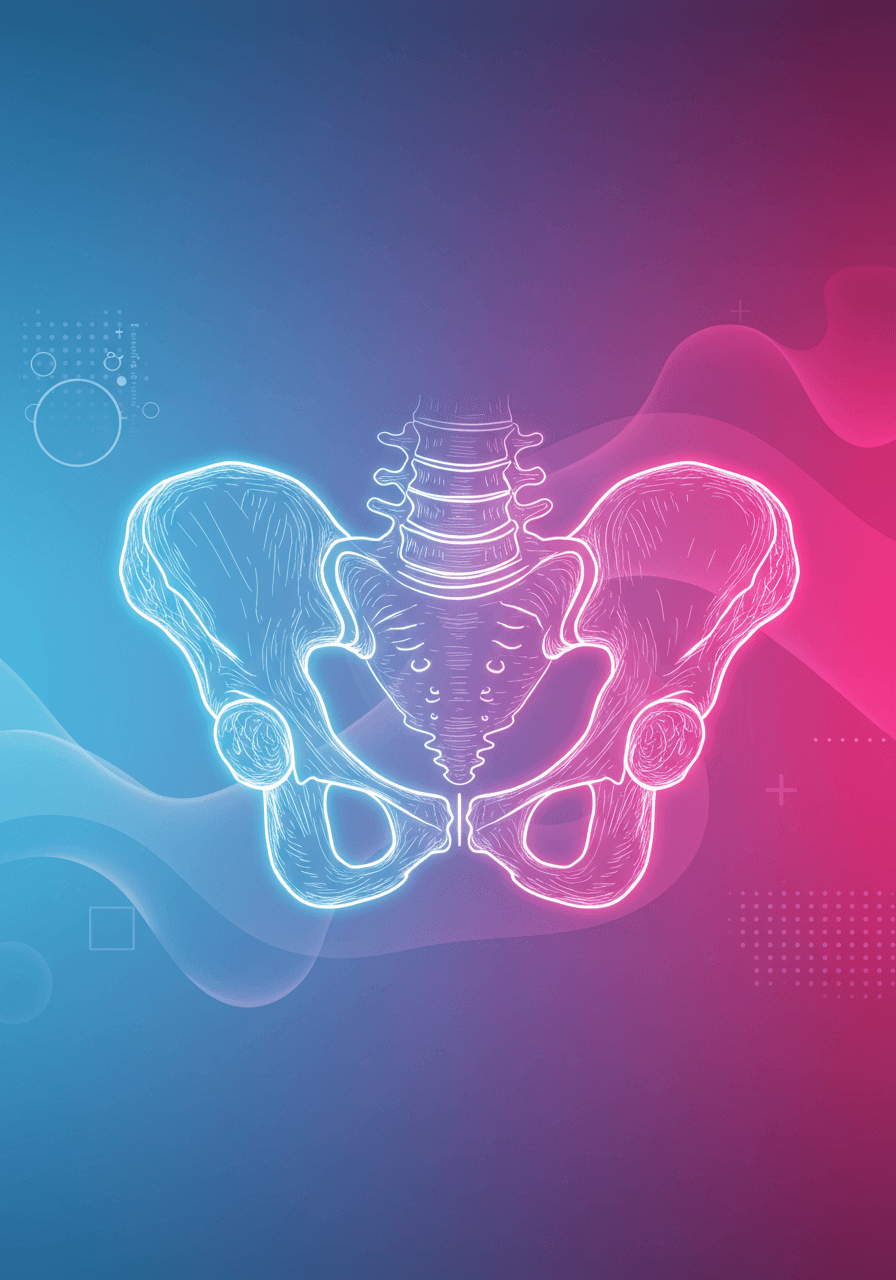
Bones of the Pelvis
- Sacrum, coccyx, and innominate bones (ilium, ischium, pubis)
- Pelvic inlet and outlet: boundaries and significance in childbirth
- Greater and lesser pelvis: boundaries and contents
Pelvic Ligaments
- Sacroiliac, sacrotuberous, and sacrospinous ligaments: attachments and functions
- Lacunar ligament and its relation to the femoral ring
Pelvic Floor Muscles
- Levator ani and coccygeus: components, attachments, and functions
- Pelvic diaphragm: role in supporting pelvic viscera
- Clinical relevance of damage to the pelvic floor muscles
Pelvic Vasculature
Arteries
- Aorta and common iliac arteries: course and major branches
- Internal iliac artery: anterior and posterior divisions and their branches
- Uterine, ovarian, vaginal, and internal pudendal arteries: course and structures supplied
- Clinical significance of the uterine artery and ureter relationship
Veins
- Internal and external iliac veins: tributaries and drainage areas
- Clinical relevance of venous plexuses surrounding the cervix and vagina
Lymphatics
- Lymphatic drainage of the pelvis: internal iliac, external iliac, and para-aortic nodes
- Clinical relevance of lymphatic drainage in malignancy
Pelvic Nerves
Lumbar Plexus
- Formation and major branches: femoral and obturator nerves
- Ilioinguinal, iliohypogastric, and genitofemoral nerves: course and clinical significance
Sacral Plexus
- Formation and major branches: sciatic and pudendal nerves
- Pudendal nerve: course, branches, and clinical relevance in childbirth
- Clinical relevance of damage to the sacral plexus
Autonomic Nervous System
- Superior and inferior hypogastric plexuses: sympathetic and parasympathetic components
- Pelvic splanchnic nerves: origin and function
- Innervation of the bladder, uterus, and rectum
IIReproductive Organs
Uterus
Structure
- Body, fundus, cervix: anatomical features and histological layers
- Internal and external os of the cervix
Ligaments
- Broad ligament: contents and subdivisions (mesometrium, mesovarium, mesosalpinx)
- Round ligament: attachments and function
- Uterosacral ligaments: attachments and function
- Transverse cervical (cardinal) ligament: attachments and function
Blood Supply and Lymphatics
- Uterine artery: course, branches, and anastomoses
- System of arteries supplying the uterus, cervix, and vagina
- Lymphatic drainage of the uterus: clinical relevance in malignancy
Vagina
Structure
- Anatomical relationships to bladder, urethra, and rectum
- Anterior and posterior fornices: clinical significance
- Histological layers of the vaginal wall
Blood Supply and Lymphatics
- Vaginal artery: origin and structures supplied
- Lymphatic drainage of the vagina: clinical relevance
Fallopian Tubes and Ovaries
Fallopian Tubes
- Parts of the fallopian tube: infundibulum, ampulla, isthmus, intramural part
- Fimbriae: structure and function
- Clinical relevance of the fallopian tubes in ectopic pregnancy
Ovaries
- Anatomical location and relationships to surrounding structures
- Suspensory ligament of the ovary (infundibulopelvic ligament): attachments
- Mesovarium: attachment to the broad ligament
- Ovarian artery: origin and course
IIIUrinary System
Bladder
Structure
- Anatomical relationships to uterus, vagina, and pubic symphysis
- Trigone: boundaries and embryological origin
- Detrusor muscle: layers and function
- Internal and external urethral sphincters
Ligaments
- Lateral true ligaments and pubovesical ligaments: attachments and function
- Median umbilical ligament: embryological remnant
Innervation
- Sympathetic and parasympathetic innervation of the bladder
- Clinical relevance of bladder innervation in urinary continence
Ureters
Course
- Course from renal pelvis to bladder, including pelvic portion
- Relationship to ovarian vessels and internal iliac artery
- Relationship to uterine artery: clinical significance during hysterectomy
Structure
- Layers of the ureteric wall
- Blood supply and innervation
Urethra
Structure
- Length and course in females
- Relationship to anterior vaginal wall
- Urethral glands and paraurethral ducts
- Histological layers of the urethra
IVPerineum and Anal Region
Perineum
Boundaries
- Boundaries of the perineum: pubic symphysis, coccyx, ischial tuberosities
- Urogenital and anal triangles: division and contents
Muscles
- Superficial perineal muscles: bulbospongiosus, ischiocavernosus, superficial transverse perineal muscles
- Deep perineal muscles: urethral sphincter, deep transverse perineal muscles
- Urogenital diaphragm: structure and function
- Perineal membrane: attachments and structures piercing it
Blood Supply and Innervation
- Internal pudendal artery: branches supplying the perineum
- Pudendal nerve: branches and areas of innervation
- Clinical relevance of the pudendal nerve in childbirth
Anal Region
Anal Canal
- Length and course of the anal canal
- Anal columns, valves, and sinuses
- Internal and external anal sphincters: structure and function
- Clinical relevance of anal sphincter injuries in childbirth
Ischioanal Fossa
- Boundaries and contents of the ischioanal fossa
- Pudendal canal: location and contents
VAnterior Abdominal Wall
Muscles
Layers
- External oblique, internal oblique, transversus abdominis, and rectus abdominis muscles: attachments, actions, and innervation
- Linea alba: formation and clinical significance
- Rectus sheath: formation and contents
- Arcuate line: significance in relation to the rectus sheath
Inguinal Region
Inguinal Ligament
- Formation and attachments
- Relationship to the inguinal canal
Inguinal Canal
- Boundaries and contents of the inguinal canal in females
- Deep and superficial inguinal rings: location and formation
- Clinical relevance of the inguinal canal in hernias
Femoral Triangle
- Boundaries and contents of the femoral triangle
- Femoral sheath: contents and clinical significance
- Clinical relevance of femoral hernias
Vessels and Nerves
Inferior Epigastric Vessels
- Origin, course, and clinical significance during laparoscopy
- Relationship to the medial and lateral umbilical ligaments
Nerves of the Anterior Abdominal Wall
- Iliohypogastric and Ilioinguinal nerves
- Genitofemoral nerve
VIBreast
Structure and Development
Anatomical Features
- Lobules, lactiferous ducts, and nipples
- Suspensory ligaments: function and clinical significance
- Areolar glands: function
Developmental Changes
- Changes in breast structure during puberty, pregnancy, and menopause
Blood Supply and Lymphatics
Arterial Supply
- Lateral thoracic artery and internal thoracic artery: branches supplying the breast
Lymphatic Drainage
- Axillary lymph nodes: clinical significance in breast cancer
- Other lymphatic drainage pathways
How to master Anatomy
- Review: work through each section systematically and understand the core concepts before moving on.
- Active learning: draw diagrams, build mind maps and explain concepts to someone else.
- Test yourself: try our free practice questions, then work through the anatomy questions in the Uterio question bank.
Additional resources
- Guidelines: RCOG Green-top and NICE guidelines give the clinical context behind anatomy questions.
- Exam logistics: check the next MRCOG Part 1 exam dates and fees and the application and booking guide.
- Essential reading: the textbooks in our revision resources list.